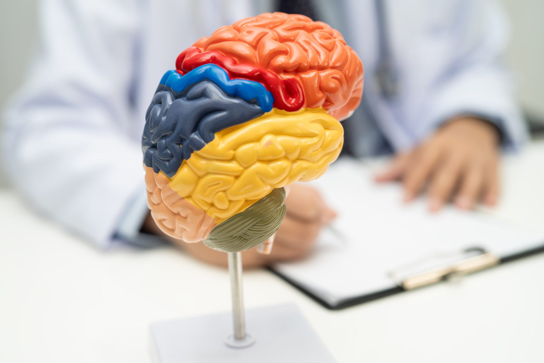
Neuro Rehab, the UK's leading event for rehabilitation professionals, brings you the latest research findings from studies conducted around the world. These studies explore various pioneering treatments for patients affected by neurological conditions or injuries, and potential new ways of improving diagnosis and long-term outcomes.
Multiple Sclerosis: Pilot study shows backward walking improves mobility in stroke patients
Researchers in the US are studying the impact of a backward walking programme on people with multiple sclerosis (MS).
A pilot study, led by Wayne State University in Michigan, found that backward walking training over eight weeks led to improvements in mobility and balance in the majority of participants.
“This suggests that backward walking may trigger positive physical adaptations,” said Dr. Nora Fritz, director of research and professor at the university’s Eugene Applebaum College of Pharmacy and Health Sciences.
The study was published in the January issue of the Journal of Neurologic Physical Therapy.
Participants took part in treadmill and overground backward walking therapies.
Dr Fritz and her team indicated additional larger clinical trials are needed to confirm the benefits.
“This novel physical therapy intervention was designed to combat the progressive movement challenges associated with MS,” said Dr Fritz.
“We measured structural changes in the brain’s white matter in three brain regions – the body of the corpus callosum, the superior cerebellar peduncle, and the corticospinal tract. The results of this small trial suggest that this type of therapy may promote neuroplasticity in brain areas related to balance. Our next step is to conduct a larger trial to determine the potential impact this type of therapy may have on all MS patients.”
Spinal cord injury: Non-invasive brain scanning could send signals to paralysed limbs
Scientists are exploring whether electroencephalography (EEG) could be a useful tool for connecting brain signals with limb movements in patients with spinal cord injuries.
When a patient tries to move their paralysed limb, their brain generates a series of signals corresponding to that movement.
But if those signals could be read and decoded, they could be relayed to a spinal cord stimulator to control nerve endings in that limb, researchers in Italy and Switzerland say.
Previous research has focused on implantable electrodes to read movement signals. While this approach has met with some success, authors wanted to study the potential of EEG devices, which are worn as caps fitted with multiple electrodes to record activity from the scalp.
“[Implantable electrodes] can cause infections; it’s another surgical procedure,” said author Laura Toni. “We were wondering whether that could be avoided.”
When electrodes are placed on the surface of a patient’s head, they struggle to pick up signals produced in the deeper regions of the brain. This poses only a small obstacle when applied to arm and hand movements but is more challenging when applied to legs and feet.
“The brain controls lower limb movements mainly in the central area, while upper limb movements are more on the outside,” said Toni. “It’s easier to have a spatial mapping of what you’re trying to decode compared to the lower limbs.”
To help them decode the EEG signals, researchers employed a machine learning algorithm designed to sift through these limited datasets.
In tests, the researchers equipped patients with EEG monitors and asked them to perform a series of simple movements. They collected the resulting data and used their algorithm to classify the range of possible signals.
They found they could detect the difference between attempted movement and no movement but struggled to differentiate between specific signals.
Going forward, researchers want to improve their algorithm to recognise different movement attempts, such as standing, walking, or climbing, and then look for ways to use that data to help trigger those movements in the implants of recovering patients.
Parkinsons: New stem cell treatment may offer hope for Parkinson’s disease
Implanting specialised stem cells into the brain could slow down the progression of Parkinson’s disease and restore motor function in patients with the condition, US scientists say.
Parkinson’s disease is associated with a reduced release of dopamine – a neurotransmitter essential for movement, memory and mood – in the brain.
Research has shown that tremors, stiffness, slow movement and other symptoms of Parkinson’s disease are caused by the progressive loss of dopamine-producing brain cells, disrupting the brain’s ability to regulate movement.
Keck Medicine of USC is conducting an early phase clinical trial investigating the safety and effectiveness of implanting lab-generated stem cells into the brain that have been programmed to replace damaged brain cells and produce dopamine.
“If the brain can once again produce normal levels of dopamine, Parkinson’s disease may be slowed down and motor function restored,” said Brian Lee, a neurosurgeon with Keck Medicine and principal investigator of the study.
The pluripotent stem cells (iPSCs) used in the study are, unlike embryonic stem cells, adult cells, reprogrammed to a "blank slate" state capable of evolving into any type of cell.
“We believe that these iPSCs can reliably mature into dopamine-producing brain cells, and offer the best chance of jump-starting the brain’s dopamine production,” said Xenos Mason, MD, co-principal investigator of the study.
During the procedure, Lee drills a small hole in the patient’s skull to access the brain, then precisely implants the stem cells into the basal ganglia, a part of the brain that controls movement, under the guidance of MRI.
After surgery, patients are monitored for 12-15 months for any changes in their Parkinson’s disease symptoms and for possible side effects including excess movements or infection. Doctors will continue to monitor the patients and their Parkinson’s disease symptoms for up to five years.
“Our ultimate goal is to pioneer a technique that can repair patients’ motor function and offer them a better quality of life,” said Lee.
Alzheimer’s Disease: Study links locus coeruleus function to deep sleep quality
A new study has reinforced the importance of deep sleep for brain health and suggest that interactions between the locus coeruleus, vascular factors, and sex may influence Alzheimer's disease progression.
The study, published in Alzheimer's & Dementia has identified that the integrity of the locus coeruleus - a brainstem structure involved in regulating the wake-sleep cycle— is associated with better deep sleep quality in healthy individuals and patients across the Alzheimer's disease spectrum.
Previous studies have shown that the status of this nucleus is associated with the severity of neuropsychiatric symptoms, including sleep problems, in people with Alzheimer’s disease. This study goes further by analysing specific deep sleep parameters.
To understand how these elements interact, the researchers at the Hospital Clínic in Barcelona analysed nighttime brain activity in 58 participants, including healthy individuals, those with mild cognitive impairment, and dementia due to Alzheimer's disease, and correlated it with markers obtained through neuroimaging techniques and biomarkers.
This approach enabled simultaneous assessment of sleep quality, locus coeruleus status, and various indicators of vascular and glymphatic health - the brain's drainage/cleaning system.
The results show that individuals with a better-preserved locus coeruleus exhibited higher slow-wave activity, an indicator of higher-quality deep sleep.
"This relationship was particularly strong in women, suggesting possible biological or hormonal differences in sleep regulation," said Neus Falgàs, first author and member of the Clínic's Alzheimer's and Other Cognitive Disorders Unit.
Additionally, a greater presence of perivascular spaces in the basal ganglia —a marker of cerebral vascular health— was associated with reduced slow-wave activity.
This combination of findings demonstrates that deep sleep quality may be influenced by both the status of sleep-regulating structures and the health of cerebral blood vessels.
The researchers say longitudinal studies are needed to determine whether preserving locus coeruleus function or improving vascular health can help maintain sleep quality and potentially modulate disease course.